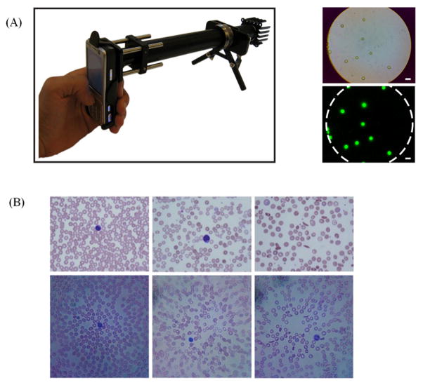
Figure 4.
(A) (Left) mobile phone microscope prototype,42 with LED and filters installed, capable of fluorescent imaging. (Right) the bright field and fluorescent imaging of 6 μm beads. (B) Micrographs of peripheral blood smears obtained by the cell phone microscope.46 Upper row: conventional microscope images. Bottom row: cell phone microscope images. Left column, images of normal blood sample. Center column, images of blood sample with iron deficiency anemia. Right column, images of blood sample with sickle cell anemia. Reprinted from references 42 and 46 with permission from PLoS One.

