Figure 6.
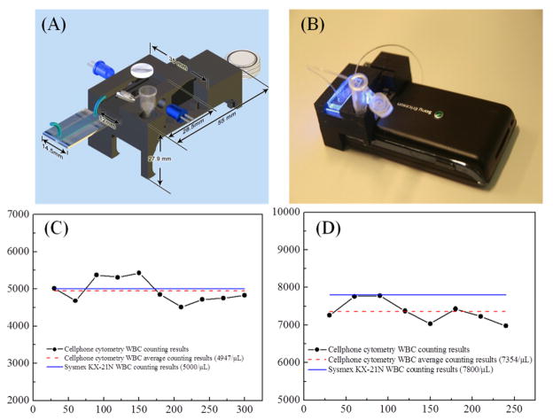
(A–B) An illustration and photograph of the fluorescent imaging flow cytometry on a cell-phone.45 The entire attachment has dimensions of ~ 35 × 55 × 27.9 mm and a weight of ~ 18 grams. Total white blood cell counting results for a low WBC density sample (5000 cells/μL) (C) and for a higher WBC density sample (7800 cells/μL) (D) obtained from the cell-phone based imaging flow-cytometer Reprinted from reference 45 with permission from ACS publishing.

