Figure 6.
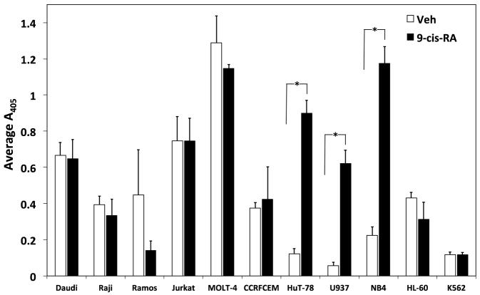
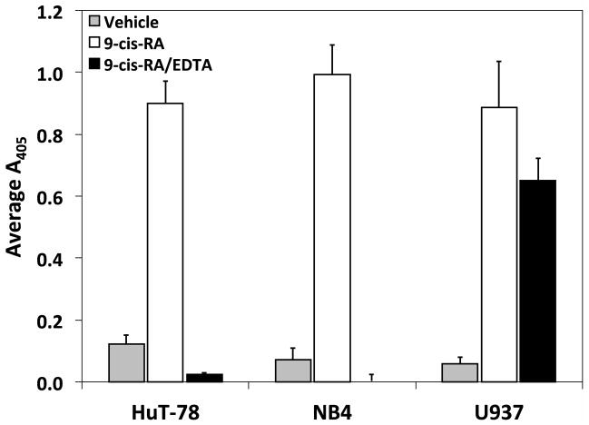
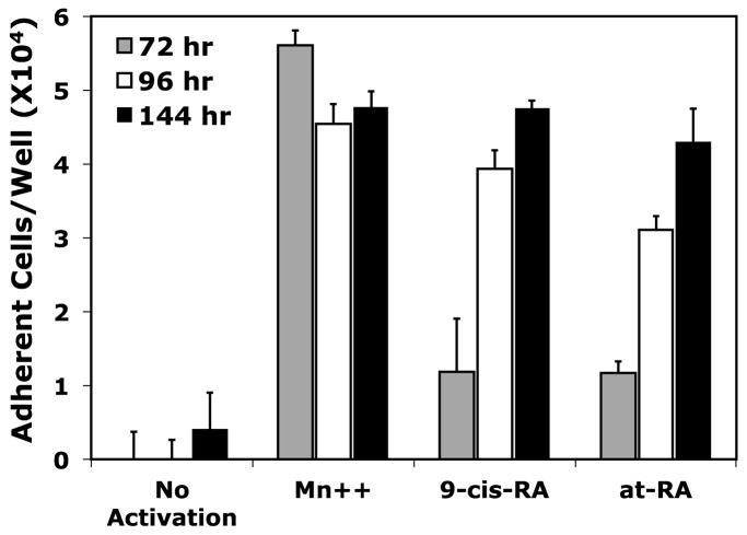
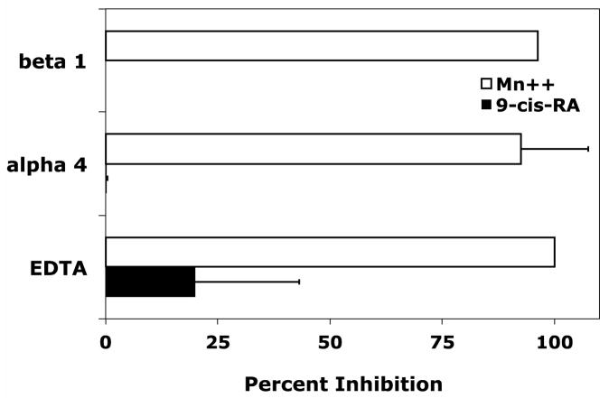
9-cis-RA dependent cell adhesion is cell specific. (A) Assorted human immortal blood cell lineages cultured for 72 hrs in the presence of vehicle (white bars) or 1 μM 9-cis-RA (black bars) were assessed for adhesion to immobilized sVCAM-Fc or ADAM28 Dis-Fc. (B) HuT-78, NB4, and U937 cells exposed to 1 μM 9-cis-RA for at least 72 hrs and subsequently evaluated for adhesion to of 3 μg/mL sVCAM-Fc in the presence or absence of 5 mM EDTA. (C) U937 cells were cultured for 72 (shaded bars), 96 (white bars) or 144 hrs (black bars) in the presence of vehicle (ethanol), or 1 μM of indicated retinoid. Adhesion assay were conducted as previously described with wells coated with 5 μg/mL of sVCAM-Fc. (D) U937 cells cultured for 96 hrs in the presence of vehicle or 1 μM 9-cis-RA were added to wells coated with 5μg/mL of sVCAM-Fc in the presence of various integrin inhibitors. Data shown are the average percent inhibition values obtained from three experiments each done in triplicate ± SD. * signifies a significant difference between adhesion values obtained with vehicle versus 9-cis-RA treated cells (Student’s t-test, p<0.01).


