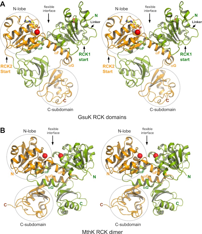
Figure 3.
Structure of the GsuK intracellular subunit. (A) Stereoviews of GsuK intracellular subunit. RCK1 and RCK2 are colored green and orange, respectively. Ca2+ and Zn2+ ions are shown as red and silver spheres, respectively. The same color representations are used in all figures. (B) Stereoviews of MthK RCK dimer. The N-terminal lobes and the C-terminal subdomains are circled in RCK2 of GsuK and in one of the RCK subunits of MthK.

