Figure 7.
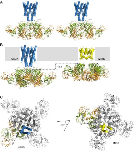
Structure of the full-length GsuK channel. (A) Stereoview of full-length GsuK channel with L97D mutation. The transmembrane helices are shown as blue cylinders and the gating ring is in ribbon representation with RCK1 in green and RCK2 in orange. Subdomains from the front and back subunits are disordered and absent in the structure. (B) Comparison of the translational distances between the gating ring and the membrane-spanning pore in GsuK (left, L97D mutant) and MthK (right). (C) Comparison of the relative orientation between the gating ring (ribbon representation) and ion conduction pore (cylinder representation) in GsuK (left) and MthK (right). Only one subunit from each channel is colored. Both channels are superimposed using the pore region and viewed from the extracellular side. Arrows connect the central fourfold axis (square) to the starting residue (Cα atoms of Gly131 in GsuK and Arg114 in MthK) of the first RCK domains, indicating the approximate direction of the linker.

