Figure 1.
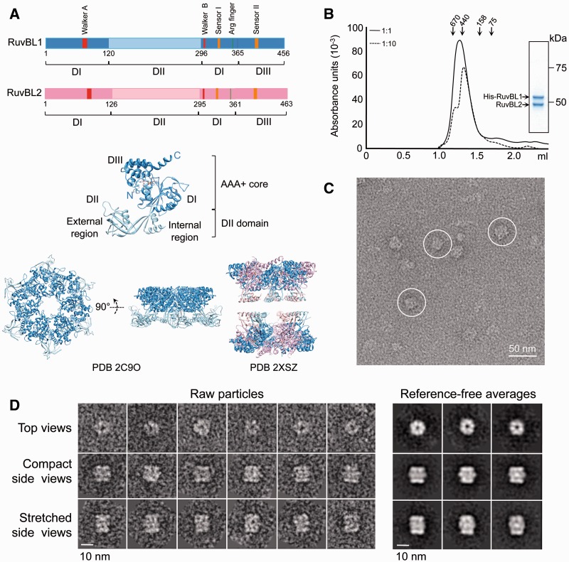
Purification and electron microscopy of human RuvBL1–RuvBL2. (A) Sequence of RuvBL1 and RuvBL2 and atomic structures of homo-hexameric RuvBL1 (PDB code: 2C9O) (16) and the truncated double-ring RuvBL1–RuvBL2 complex (PDB code: 2XSZ) (17). The middle panel shows an enlarged view of a RuvBL1 monomer. Domains and the N- and C-terminal ends of the protein are labelled. Colour codes for different domains in the primary structure are used in the atomic structures. (B) Elution profile from a SEC of the His–RuvBL1–RuvBL2 complex after affinity purification (solid line). The sample was also analysed after 1:10 dilution (dashed line). Molecular weight standards corresponding to 670, 440, 158 and 75 kDa are indicated. Inset shows a SimplyBlue (Novagen) stained SDS–PAGE of purified RuvBL1–RuvBL2 complex after SEC. (C) Representative field of an electron micrograph obtained for RuvBL1–RuvBL2 after negative staining. Selected side view images are highlighted. Scale bar, 50 nm. (D) Raw images and reference-free 2D averages obtained after reference-free classification and averaging. Scale bar, 10 nm.

