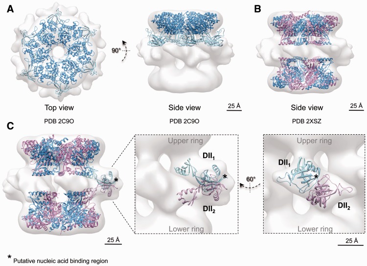
Figure 6.
Cryo-EM structure and pseudo-atomic model of the compact conformation of human RuvBL1–RuvBL2. (A) Top and side views showing the fitting of the atomic structure of RuvBL1 (PDB code: 2C9O) (16) in the top ring. (B) Fitting of the atomic structure of the truncated human RuvBL1–RuvBL2 complex (PDB code: 2XSZ) (17). RuvBL1 is shown in blue and RuvBL2 in pink. Scale bar, 2.5 nm. (C) DII domains were fitted into the density of the cryo-EM map assigned to these domains after splitting external and internal regions. An asterisk points to putative regions of DII domains involved in the interaction with nucleic acids, hypothesized based on the comparison with RPA (see text). Scale bar, 2.5 nm.

