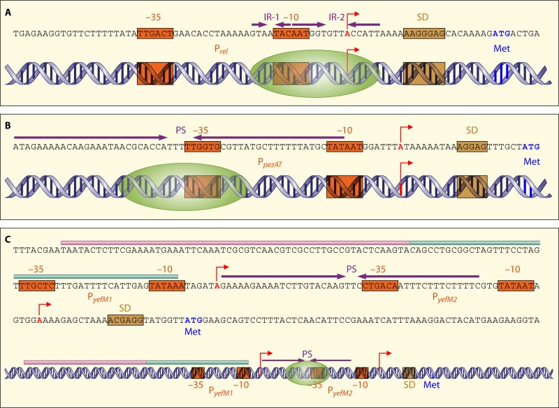
Fig 3.
Schematic representation of the DNA double helix at the promoter regions of the pneumococcal relBE2 (A), pezAT (B), and yefM-yoeB (C) TAS as well as the (putative) binding sites of their respective proteins. The inverted repeats (IR) or palindrome sequences (PS) are indicated by purple arrows, the transcriptional start sites are indicated by red arrows, and the boxA and boxC subelements are indicated by pink and green lines, respectively. The −10 and −35 promoter sequences (orange), the Shine-Dalgarno (SD) sequences (brown), as well as the start codon “Met” (navy blue) are also indicated. The proposed binding sites of the TA complexes (green ovals indicate the TA proteins) are also shown.

