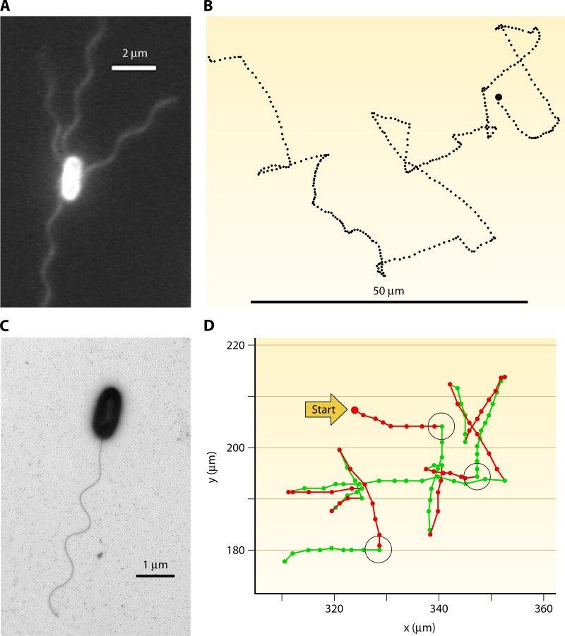
Fig 7.
Flagellation and motility patterns, illustrating fundamental differences between enteric model organisms and some marine bacteria. (A) Epifluorescent image of Escherichia coli, showing multiple flagella. Reproduced from reference 194 with permission. (B) E. coli swims in a run-and-tumble pattern, where each nearly straight swimming segment (run) is interrupted by a nearly random change in direction (tumble). The image shows a 30-s trajectory containing 26 runs and tumbles. The trajectory spans ∼100 μm from left to right. (Reproduced from reference 25a by permission from Macmillan Publishers Ltd. Copyright 1972.) (C) Transmission electron microscopy image of Vibrio alginolyticus, showing the single polar flagellum (K. Son, J. S. Guasto, and R. Stocker, unpublished). (D) The “hybrid” swimming pattern of Vibrio alginolyticus, alternating reversals (180-degree changes in directions) after each forward run (green segments) and large reorientations after each backward run (red segments). The reorientations, some of which are highlighted by black circles, are caused by a whip-like deformation of the flagellum (a “flick”). Dots are 1/30th of a second apart. (Reproduced from reference 207 with permission.)

