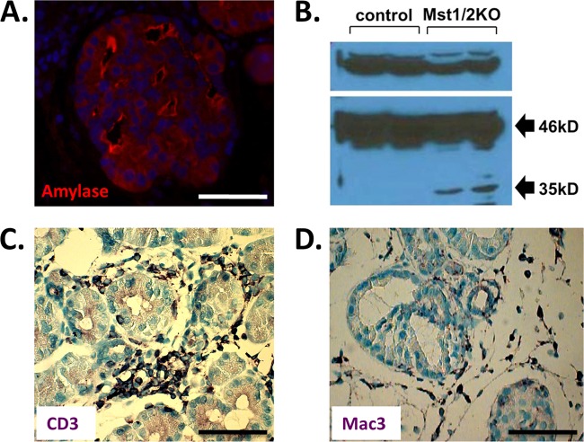
Fig 5.
Pancreas autodigestion counteracts cell proliferation. (A) Transitional structures display intense amylase staining along the cell surface of duct-like lumens suggestive of tissue autodigestion. (B) In contrast to controls, 6-week Mst1/2 KOs display intrapancreatic activation of carboxypeptidase A (CPA). Only the 46-kDa proform of CPA is evident in controls, whereas both the proform and the 35-kDa catalytically active form are present in the knockouts. (C and D) Mst1/2 KOs display a robust inflammatory infiltrate consisting of lymphocytes (e.g., CD3+ cells) and macrophage (Mac3+). Scale bar, 50 μm.

