
Fig 4.
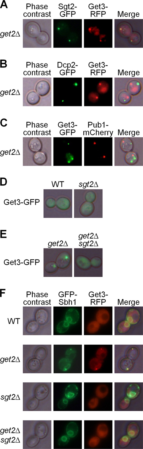
Aggregation patterns in strains with the get2Δ and/or sgt2Δ deletion. (A) In the get2Δ strain, Get3 (tagged by red fluorescent protein [RFP]) and Sgt2 (tagged by GFP) formed aggregates that were colocalized. (B and C) Get-TA aggregates, marked by fluorescently tagged Get3, did not colocalize with the P-body marker Dcp2 (B) or with the stress granule marker Pub1 (C). (D) The sgt2Δ deletion did not cause the formation of Get-TA aggregates (marked by Get3-GFP) in the Get+ strain. (E) The sgt2Δ deletion abolished aggregation of the cytosolic Get protein Get3-GFP in the get2Δ strains. (F) The tail-anchored protein Sbh1 (tagged by GFP) was localized to the endoplasmic reticulum membrane in the wild-type (WT) strain but was colocalized with the Get3 aggregates in the get2Δ strain. Membrane localization of Sbh1 was not altered by the sgt2Δ deletion and was restored in the get2Δ sgt2Δ strain.