
Fig 1.
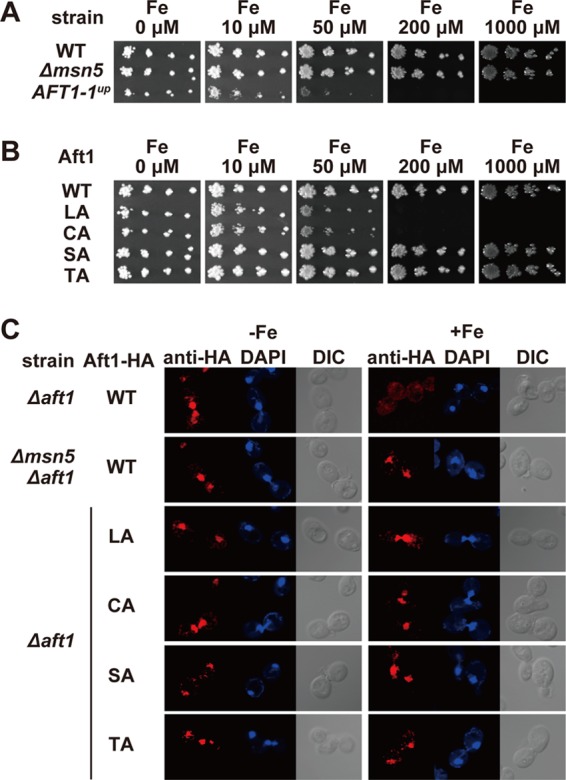
The Δmsn5 strain is not sensitive to excess iron. (A) The indicated strains were spotted on SD medium supplemented with the indicated concentrations of FeSO4 using 3-fold serial dilutions beginning at 200 cells per spot. Cell growth was observed after incubation at 30°C for 3 days. (B) The growth of cells expressing the indicated Aft1p mutants was tested as for panel A. (C) Aft1p accumulates in the nucleus of the Δmsn5 strain independent of cellular iron status. Δaft1 or Δmsn5 Δaft1 cells carrying an expression plasmid for wild-type Aft1-HA or the indicated mutants were cultured in iron-depleted (−Fe) or iron-replete (+Fe) medium to mid-log-phase growth. After fixation, the subcellular localization of Aft1-HA was examined by indirect immunofluorescence microscopy using an anti-HA antibody. DAPI staining of nuclei and differential interference contrast (DIC) images are provided for comparison.