Fig 5.
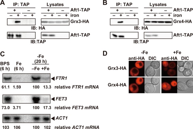
The Grx3/4p interaction with Aft1p is augmented in the presence of iron. (A and B) Binding of Grx3/4p to Aft1p is enhanced under iron-replete conditions. (A) Cells expressing both Aft1-TAP and Grx3-HA (+ Aft1-TAP) or Grx3-HA alone (− Aft1-TAP) from their natural chromosomal loci were cultured in iron-free medium to mid-log-phase growth (20 h). Cells were cultured for an additional 15 min in the absence (− iron) or presence (+ iron) of 200 μM FeSO4, and TAP immunoprecipitates and cell lysates were probed with the indicated antibodies. (B) Cells expressing both Aft1-TAP and Grx4-HA (+ Aft1-TAP) or Grx4-HA alone (− Aft1-TAP) from their natural chromosomal loci were cultured as for panel A, and the interaction between Aft1-TAP and Grx4-HA was analyzed by immunoprecipitation. (C) Induction of the expression of the iron regulon is incomplete after a 6-h treatment with 40 μM BPS. BY4741 cells were cultured under the following conditions: SD medium containing 40 μM BPS for 6 h (BPS), SD medium containing 250 μM FeSO4 for 6 h (Fe), SD medium lacking iron for 20 h (Fe; −), or SD medium lacking iron for 20 h with an additional 15-min cultivation in the presence of 200 μM FeSO4 (Fe; +). Total RNA was extracted, and expression of the indicated genes was analyzed by Northern blotting. The values indicated below each panel are relative mRNA levels as a percentage of the amount in the cells cultured in SD medium lacking iron for 20 h. (D) Grx3/4p reside both in the nucleus and in the cytoplasm. Δgrx3 Δgrx4 cells carrying expression plasmids for Grx3/4-HA were cultured in iron-free medium to mid-log-phase growth. Cells were cultured for an additional 30 min in the absence (−Fe) or presence (+Fe) of 200 μM FeSO4. After fixation, the subcellular localization of Grx3/4-HA was examined by indirect immunofluorescence microscopy using an anti-HA antibody. Differential interference contrast (DIC) images are provided for comparison.

