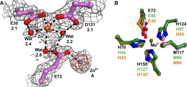
Fig 4.
Metal binding in Tp34 at site α and a comparison of sites A from three proteins. (A) Metal binding at site α in Tp34. The color, map, and notation conventions are as described in the legend to Fig. 3. The site α Zn2+ is shown as an orange sphere. Anomalous map contour = 15σ. (B) Sites A from three proteins. Shown are residues from Tp34 with Zn2+ bound (colored as in Fig. 2), CJp19 (orange carbon atoms), and FetP (green carbon atoms). The metal ions from CJp19 and FetP are coppers; they are colored the same as the carbon atoms from their respective structures. The superpositions were performed using all Cα atoms from the respective proteins. The residue numbers are noted; those belonging to Tp34 are black, while those belonging to the other two structures are colored according to their respective carbon atom colors.

