
Fig 1.
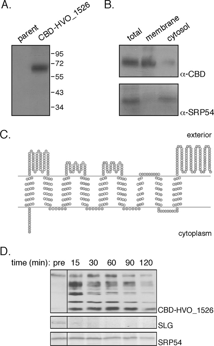
Hfx. volcanii HVO_1526 is a membrane protein. (A) Total extracts of Hfx. volcanii parent strain cells and cells transformed to express CBD-HVO_1526 were probed with anti-CBD antibodies. The positions of molecular weight markers are indicated on the right. (B) Hfx. volcanii cells transformed to express CBD-HVO_1526 were separated into membrane and cytosolic fractions and probed with anti-CBD (α-CBD) and anti-SRP54 (α-SRP54) antibodies, as was a total protein extract. (C) Schematic depiction of the topology of HVO_1526 according to the SOSUI server, drawn using Topo2 software (http://www.sacs.ucsf.edu/TOPO2/). The N terminus is found at the left end of the sequence. (D) Hfx. volcanii cells transformed to express CBD-HVO_1526 were challenged with proteinase K (1 mg/ml; 2 h at 55°C). After separation by SDS-PAGE, the S-layer glycoprotein (SLG) was identified by Coomassie staining (middle panel), while CBD-HVO_1526 (upper panel) and SRP54 (lower panel) were identified by immunoblotting using appropriate antibodies. The presence of CBD-HVO_1526 (and derived proteolytic fragments), the S-layer glycoprotein, and SRP54 was assessed before the addition of proteinase K (pre) and 15, 30, 60, 90, and 120 min after proteinase K addition.