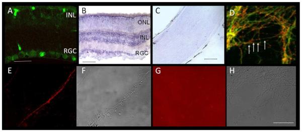
Figure 4. hnRNP-M is expressed in RGCs.
(A) RGCs and cells in the INL label with anti-hnRNP-M antibody. (B) In situ hybridization demonstrates strong labeling of hnRNP-M mRNA in RGCs and the INL (dark purple) in the retina, consistent with antibody labeling. (C) In situ hybridization in optic nerve does not detect hnRNP-M mRNA in cell bodies endogenous to the nerve. (D) In RGC explant cultures in vitro, hnRNP-M (red) localizes to axons of RGCs (green) in a punctate pattern (arrows). (E) Labeling unfixed, live cultures with an anti hnRNP-M antibody to an epitope that corresponds to the ligand binding region of rat hnRNP-M demonstrates that hnRNP-M is expressed in RGC axons and localizes to the cell surface (red). (F) Non-fluorescent image of (E). (G, H) Secondary-only control confirms that the fluorescence labeling in E is due to anti hnRNP-M binding the cell surface. INL=inner nuclear layer, RGC=retinal ganglion cell layer, ONL=outer nuclear layer. Scale bars as follows: A=20 μm; B=100 μm; C, D-H=50 μm.

