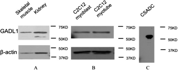
FIGURE 5.

Western blot analysis of GADL1 in tissues and cell culture. A, the presence of GADL1 in mouse kidneys and muscles. Noticed that a lower band was detected in kidneys; this band corresponded to CSADC. B, the presence of GADL1 in mouse C2C12 myoblasts and myotubes. C, 10 μg of purified mouse CSADC was used to detect whether the GADL1 antibody had cross-reactivity. Antibody against β-actin was used as a control.
