FIGURE 3.
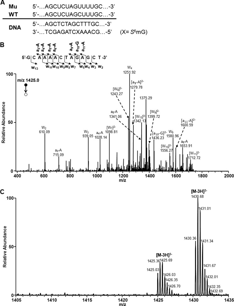
LC-MS and MS/MS for monitoring the 14-mer restriction fragments of S6mG-bearing substrate from the in vitro transcription with T7 RNAP. A, the sequences of WT and mutant (Mu) transcripts are indicated above the double-stranded DNA construct. B, product ion spectrum (MS/MS) of the [M-3H]3− ion (m/z 1425.0) of the complementary 14-mer fragment of the mutant sequence d(GCAAAACTAGAGCT). Shown above the spectrum is a scheme summarizing the observed [an − Base] and wn fragment ions, and nomenclature follows that described previously (55). C, high resolution “ultra zoom-scan” ESI-MS revealed the presence of the [M-3H]3− ions of the wild-type sequence d(GCAAAGCTAGAGCT) (m/z 1430.4) and the mutant sequence d(GCAAAACTAGAGCT) (m/z 1425.0) but the absence of the [M-3H]3− ions of other single-base substitution products of d(GCAAAMCTAGAGCT) (M = C or T, with the calculated m/z being 1417.2 and 1422.2, respectively).

