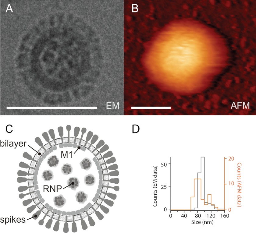
FIGURE 1.
Imaging using AFM does not deform the virus particles. Shown are a cryo-electron micrograph (A) and AFM scan (dynamic mode) (B) of a native A/X-31 influenza virus. Scale bars, 100 nm. Whereas spike glycoproteins, HA, and NA could be identified by cryo-electron microscopy, the particles imaged by AFM appeared smooth. The lateral dimensions of the AFM scans are exaggerated due to tip-sample dilation. C, schematic representation of the influenza virus structure. The viral ribonucleoprotein core (RNP) inside the virus particle is surrounded by a lipid bilayer with associated matrix protein M1 interacting with the cytoplasmic tails of spike glycoproteins HA and NA. D, histograms of the diameter of single virus particles imaged by cryo-EM and measured by atomic force microscopy (height measurement, not affected by tip-sample dilation).

