FIGURE 3.
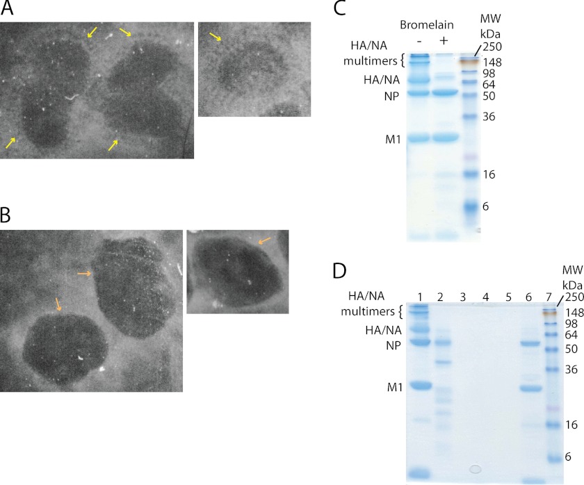
A, negative stain electron micrographs of WT influenza virions. The yellow arrows mark the ectodomains of the glycoproteins protruding from the particle surface. B, negative stain electron micrographs of WT virions after bromelain treatment. The ectodomains of the glycoproteins are no longer visible, and the surface of the particles (orange arrows) appears smooth. C, bromelain-treated A/X31 virus samples were analyzed by 12.5% SDS-PAGE in non-reducing conditions. Gels were stained with Coomassie Blue. D, 12.5% SDS-PAGE in non-reducing conditions. Lane 1, untreated virus; lane 2, supernatant of bromelain-treated sample after ultracentrifugation; lanes 3–5, three pellet washes following bromelain treatment and ultracentrifugation; lane 6, pellet of bromelain-treated virus after ultracentrifugation.

