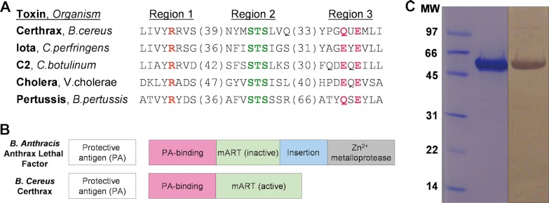
FIGURE 1.
In silico identification of Certhrax as a mono-ADP-ribosyltransferase. A, sequence alignment of Certhrax with several other mART toxins. Characteristic active site residues are shown to be conserved in region 1 (catalytic arginine, orange), region 2 (STS motif, green), and region 3 (catalytic QXE/EXE motif, pink). B, domain organization of B. anthracis anthrax LF (top) as compared with B. cereus Certhrax (bottom). Each toxin contains a separate PA protein (white) and an enzyme component (gray) which includes a PA-binding domain (pink) and a mART domain (green). Anthrax LF includes an additional insert domain (blue) and a zinc metalloprotease domain. C, SDS-PAGE gel and anti-His tag Western blot showing purified Certhrax. Lane 1, molecular mass standards (Bio-Rad) in kDa; lane 2, purified Certhrax; lane 3, Western blot, purified Certhrax.

