FIGURE 1.
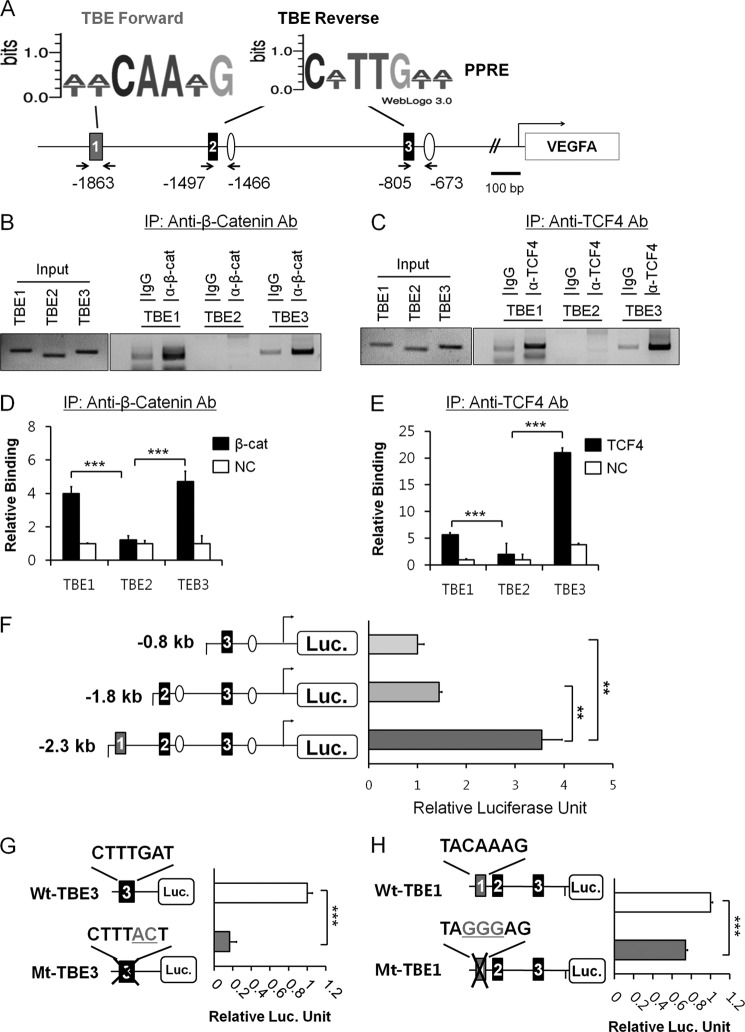
Mapping of upstream TBEs for β-catenin-mediated VEGFA transcription. A, shown is the predicted TBE forward, TBE reverse and PPAR-response elements (PPREs) in the upstream of VEGFA gene. Numbers in the boxes denote TBE sites from the upstream. 2 kb upstream to 1 kb downstream from the transcription start were analyzed for the predicted sites. Consensus sequences of the predicted TBEs in forward and reverse orientation are also shown by WebLogo3.0. B, ChIP analysis of TBE1, TBE2, and TBE3 in HCT116 colorectal carcinoma cells were grown in serum-free basal condition. Anti-β-catenin antibody (Ab) was employed in addition to IgG control antiserum. Input DNA was also shown to confirm the equal loading of the samples. IP, immunoprecipitate. C, ChIP analysis of TBE1, TBE2, and TBE3 with anti-TCF-4 antibody was as in B. D, ChIP-qPCR analysis for the TBEs using anti-β-catenin antibody is shown. Bound DNA was analyzed by quantitative real-time PCR and is presented as relative percent input of chromatin. Three independent analyses were performed; the results represent the average ± S.D. (***, p < 0.005). NC denotes negative control without antibody. E, ChIP-qPCR analysis for the TBEs using anti-TCF-4 antibody is shown. Bound DNA was analyzed by quantitative real-time PCR as in D. Three independent analyses were performed; the results represent the average ± S.D. (***, p < 0.005). F, a luciferase assay with various deletions in the upstream regions of VEGFA gene is shown. pGL2-VEGFA promoter reporters have −2.3 kb (from TBE1), −1.9 kb (from TBE2), and −0.8 kb (from TBE3) upstream fragment of VEGFA gene. HCT116 cells were transiently transfected with pGL2-VEGFA promoter reporter and pRL-TK and GW501516 treatment. Three independent experiments were performed, and the results represent the average ± S.D. (**, p < 0.01). G, shown is a luciferase assay with wild-type (Wt-TBE3) or mutant TBE3 sequences (Mt-TBE3) in the VEGFA promoter. The experiment was performed as in F and is shown in relative luciferase activities in comparison to that of wild type. Three independent experiments were performed; the results represent the average ± S.D. (***, p < 0.005). H, shown is a luciferase reporter assay with wild-type (Wt-TBE1) or mutant TBE1 site (Mt-TBE1) in the VEGFA promoter. The experiment was performed as in F and shown in relative luciferase activities in comparison to that of wild type. Three independent experiments were performed; the results represent the average ± S.D. (***, p < 0.005).

