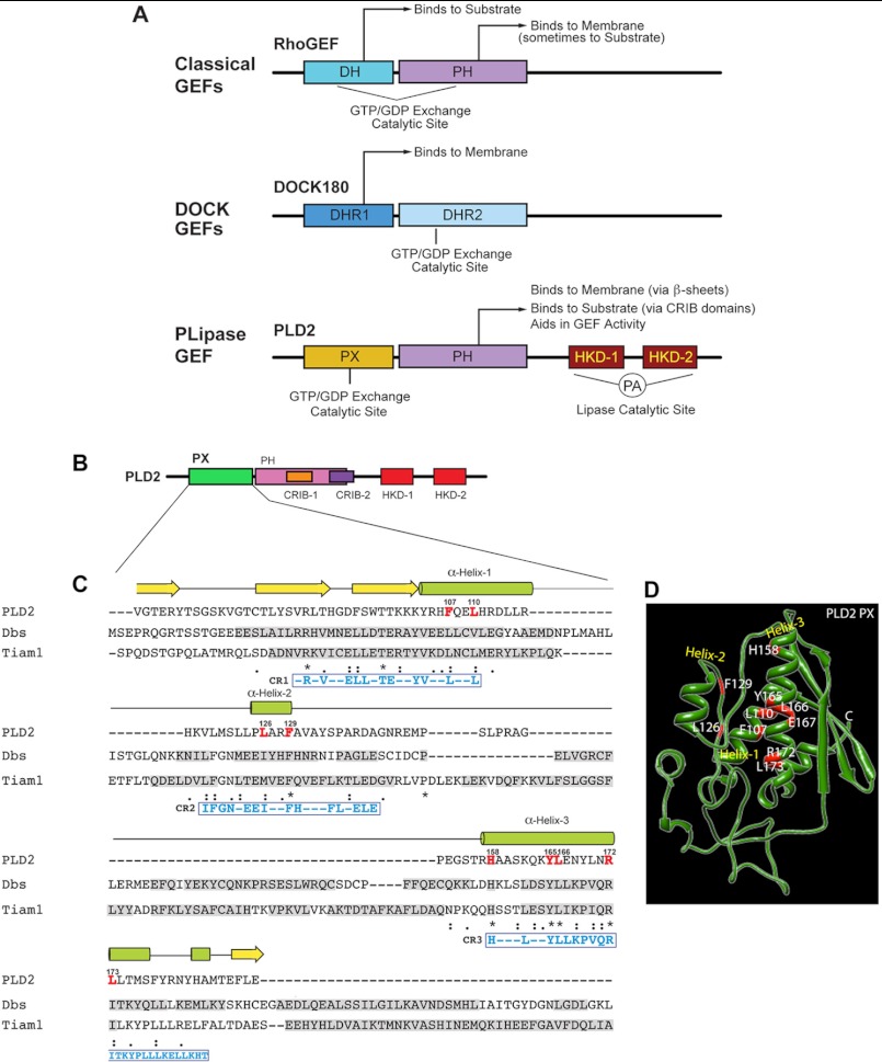
FIGURE 4.
Sequence alignments of PX in PLD2 with DH in Dbs and Tiam1. A, shown are schemes depicting the major classes of Rho GEFs with their catalytic GEF domains, as well as the newly-defined PLD2-GEF in this study. B, shown is a schematic representation of PLD2 with the PX domain highlighted in green. C, sequence alignments of PLD2 PX, Dbs DH and Tiam1 DH domains were performed using ClustalW (29). α-Helices and β-sheets of PLD2 are shown on the top of the sequence in green and yellow, respectively. Sequences of α-helices and β-sheets of Dbs and Tiam1 are shown shaded in gray within their respective sequences. Residues conserved within the DH domains of Dbs and Tiam1 and within the helices of the PLD2 PX domain are highlighted in red letters. Identical amino acids, similar amino acids and nearly similar amino acids are shown underneath the sequences as stars, as a single dot, or as double dots respectively. D, shown is a predicted three-dimensional structure schematic of PLD2 PX generated by I-TASSER. Also shown are the mutations considered in this study.

