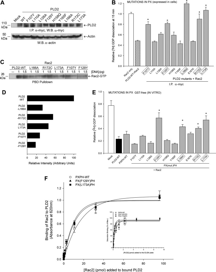
FIGURE 5.
Effect of mutations in the PX domain on PLD2 GEF activity. The PX point mutations were made in pcDNA3.1-mycPLD2 (full-length). Panel A shows protein expression levels of the PLD2 GEF mutants generated for this study using Western blot (W.B.) analysis and immunoprecipitation (I.P.). B, the effect of single point mutations of the PLD2 PX domain on Rac2 GDP dissociation was determined by two approaches. PLD2 residues involved in GEF activity in whole cells are outlined in gray boxes. We called this GEF activity in whole cells because PLD2 was overexpressed and immunoprecipitated (I.P.) from COS-7 cells (first approach). Activity of I.P.-PLD2WT or mutants was determined using recombinant Rac2 as the substrate. C, in vivo Rac2 activation (PBD pulldown) using PLD2-WT and relevant PLD2 mutants overexpressed in cells. D, horizontal bar graph representing relative intensity of Rac2-GTP in panel C. E, Rac2 GDP dissociation in the presence of purified recombinant GST-free PXPH (second approach) with mutations in PX domain is shown. Error bars are S.E., and significant (p < 0.05) differences with controls are shown by asterisk (*). Residues found to affect GEF activity using purified, recombinant proteins are outlined in gray boxes. F, PLD2 Rac2 binding is shown. The inset indicates that the PXPH-WT or mutants were bound to ELISA plates until saturation (gray ellipse). Increasing amounts of baculoviral, purified HA-Rac2 (0.043–96.15 pmol) were laid on top of the PXPH-coated wells, and bound Rac2 was detected with specific antibody. Protein binding was measured by spectrometry at A620 nm. Results represent the mean ± S.E. for four independent experiments.

