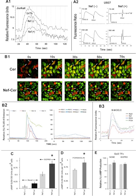
FIGURE 2.
Nef markedly inhibited biochemical readouts of Gαi2 activation. Nef inhibits chemoattractant-mediated calcium flux in Jurkat cells (A1) and the U937 cell line (A2). Cells were co-transfected with CD8 and Nef, co-expressers were purified by CD8-positive selection (Stem Cell Technologies), and Nef effect in Jurkat cells was evaluated by measuring cell surface CD4 expression. The time course of CXCL12 (10 nm)-initiated calcium flux profiles was obtained using FlexStation III and the recommended assay. Results are representative of four experiments. Agonist (CXCL12 followed by CCL2) dose (10 or 100 nm)-response profiles of intracellular calcium flux in U937 transfectants were analyzed by fluorescence ratiometry in a PTI fluorimeter (33). B1, time course of CXCL12-initiated Ca2+ flux in HeLa cells expressing CerFP or Nef-CerFP (red) was monitored by video capture at 30 frames/s of Fluo-4 emission (green) up to 150 s after CXCL12 addition. The arrows denote CerFP or Nef-CerFP cells (expressed at ∼10–15% efficiency around 12–16 h post-transfection) to highlight their difference in Fluo-4 intensity. Fluorescent data were collected from ∼10 ROIs for each field, the calcium flux was measured in >5 fields for each condition in an experiment, and the experiments were repeated three times (n = 50–60). The change in the intensities was analyzed using the Leica software followed by graphing using EXCEL. Ca2+ flux profiles of a few (to avoid clutter) representative cells (ROIs) expressing CerFP (left) or Nef-CerFP (right) are shown in B2, with the ordinate showing relative intensity of Fluo-4 emission. CXCL12-initiated Ca2+ flux is profiled in purified CEM cells co-transfected with CD8 and WT, null, or other Nef mutants. CEM cells were transfected with CD8, WT, null, or the indicated Nef mutants, and CD8(+) cells were purified prior to measurement of Ca2+ flux (as described above) (B3). Nef expression enhanced cAMP levels under basal conditions or after Gαs activation by isoproterenol (C) or by forskolin stimulation of adenylyl cyclase (D). However, Nef did not further enhance the cAMP levels after isoproterenol treatment with transfectants overexpressing Gαi3 (E). Jurkat cells were cotransfected with CD8 and Nef or vector (for C–E) and with a Gαi3 expression plasmid (only for E). Transfected cells were purified by CD8 selection and assayed for cAMP production as described under “Experimental Procedures” (n = 4; error bars represent S.E.; *, p < 0.05).

