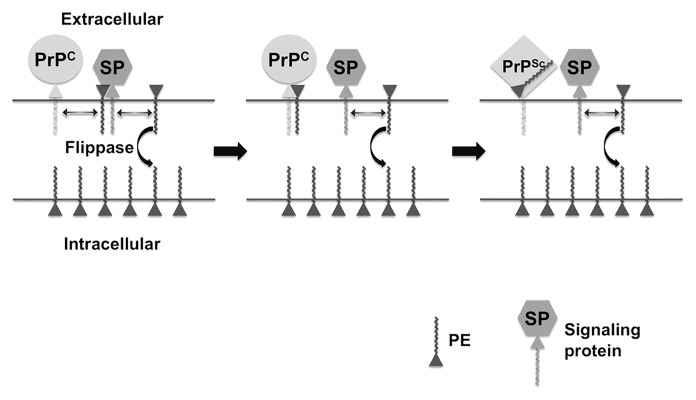
Figure 1. Hypothetical model of PE in prion formation and pathogenesis. Prion Formation: the relative paucity of PE on the extracellular leaflet of the plasma membrane of cells is actively maintained by a flippase enzyme, whose activity declines with age, allowing more PE to be localized on the extracellular surface where it is accessible to PrPC. Pathogenesis: If extracellular PE becomes consumed during the process of prion formation by binding irreversibly to PrPSc, its depletion from the membrane surface will cause biophysical changes in the membrane itself and dysregulation of surface-expressed signaling proteins (SP) that normally bind PE, resulting in cell death.

An official website of the United States government
Here's how you know
Official websites use .gov
A
.gov website belongs to an official
government organization in the United States.
Secure .gov websites use HTTPS
A lock (
) or https:// means you've safely
connected to the .gov website. Share sensitive
information only on official, secure websites.
