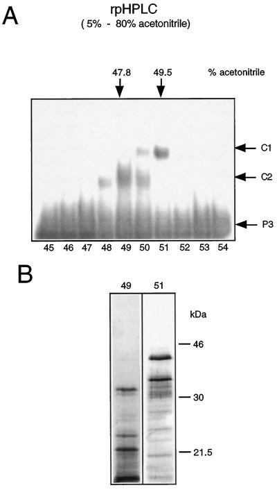
Figure 3.
Separation of DNA-binding proteins by rpHPLC. A, Phosphocellulose fractions with binding activities (Fig. 1C, fraction nos. 6–20) were pooled and chromatographed on an rpHPLC C4 column. Fractions eluted with a linear acetonitrile gradient (5–80%) were tested after renaturation for DNA-binding activity by mobility-shift analysis using ds-oligonucleotide P3. Arrows in the right margin indicate the positions of the DNA-protein complexes C1 and C2 and of the unbound ds-oligonucleotide P3. Fraction numbers are given at the bottom. B, SDS-PAGE analysis of rpHPLC fraction nos. 49 and 51. Proteins were resolved on 12.5% SDS-polyacrylamide gels and visualized by silver staining. Molecular masses of marker proteins are given in kilodaltons.

