Figure 2.

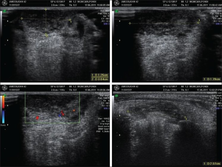
Ultrasound color Doppler study showing hypo echoic with multiple tiny cystic areas and mild internal vascularity within the lesion

Ultrasound color Doppler study showing hypo echoic with multiple tiny cystic areas and mild internal vascularity within the lesion