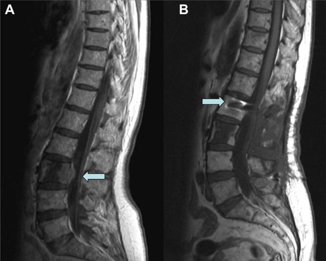
Figure 1.
A preoperative lumbosacral MRI showed a L3 involvement by lymphomatous tissue with evidence of intracanalar diffusion as indicated by arrow (A). A postoperative MRI with evidence of surgical procedure of decompressive laminectomy and stabilization with titanium pedicle screws (see arrow) and rods (B).

