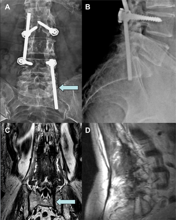
Figure 3.
AP and LL lumbar x-ray performed after an S1 left radiculopathy onset showed the migration of both paravertebral rods, with the left one migrated in the pre-sacral region and the right one positioned at the upper level of second lumbar vertebral body (A and B). A coronal and sagittal plane of preoperative MRI allowed the visualization of the bone sacral groove created by the migrated left rod as marked by arrows (C and D).

