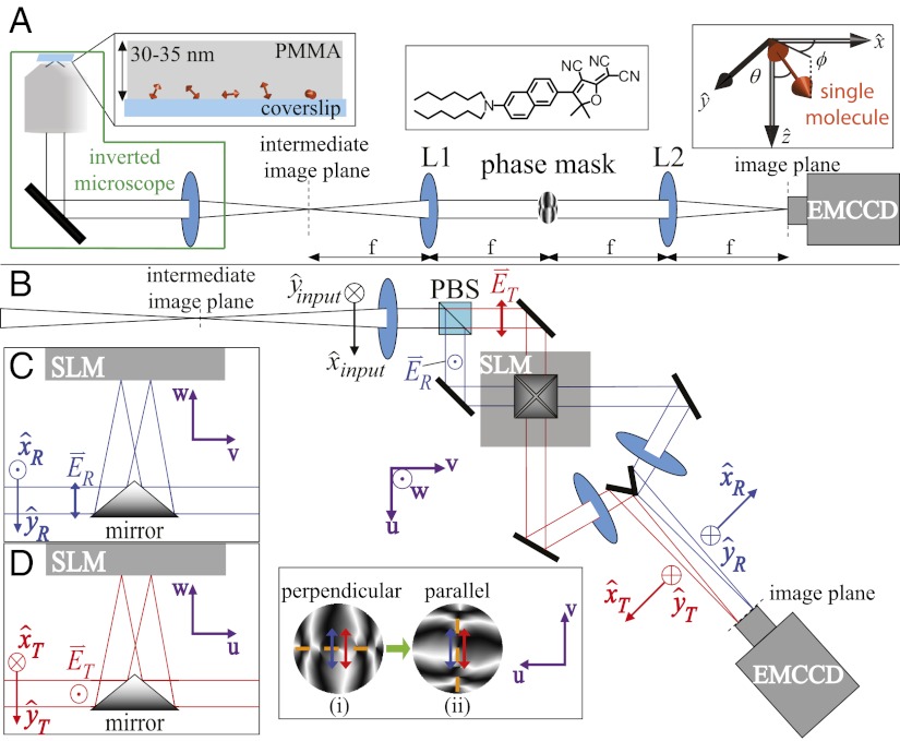
Fig. 1.
DH-PSF imaging system. (A) Inverted microscope and 4f optical system schematic, where L1 and L2 are focal length-matched achromatic lenses. Our sample for the experiments described consisted of DCDHF-N-6 molecules (Center Inset) embedded in a thin layer of PMMA (Left Inset). Orientation angles (θ, ϕ) are defined in Right Inset and have ranges (0°, 90°) and (−180°, 180°), respectively. (B) The high efficiency dual-polarization detection DH-PSF setup used for these experiments (inverted microscope omitted for simplicity). The collected fluorescence is split by a polarizing beam splitter (PBS) into reflected (R; blue) and transmitted (T; red) channels. Input Cartesian unit vectors  define molecular orientation (θ, ϕ) and are propagated differently through the various reflections in the two polarization channels [
 and
and  ]. The two electric field polarization axes
  and
and  are projected identically onto the phase mask (Inset). Inset shows how each polarization axis (blue and red arrows) is oriented relative to the mask’s axis of phase discontinuities (dashed orange) when the mask is upright (i; polarization perpendicular to discontinuities) and rotated (ii; polarization parallel to discontinuities). (C and D) Two side-on views of the SLM portion of the setup showing the square pyramidal mirror.

