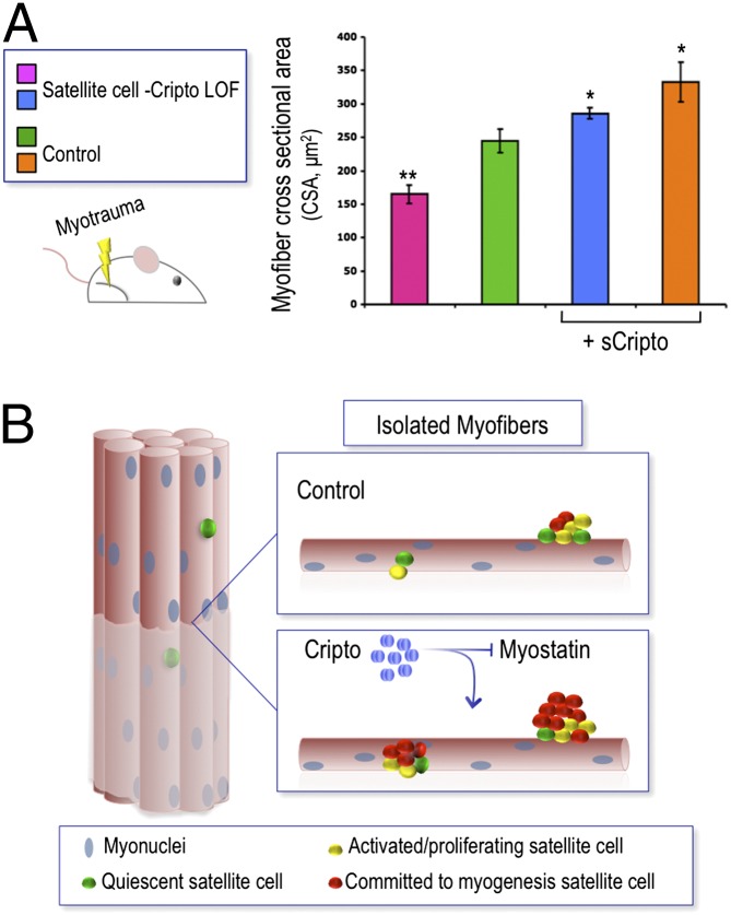
Fig. P1.
(A) (Left) Schematic representation of a mouse model of skeletal muscle regeneration induced by acute injury. LOF, loss of function. (Right) Myofiber cross-sectional area (CSA) analysis of skeletal muscles in different experimental conditions shows that muscle regeneration is affected when satellite cells lose Cripto function (satellite cell Cripto LOF, pink bar) compared with control (green bar) (mean ± SEM; n = 3 mice/group; *P = 0.02; **P < 0.004). Muscle regeneration defects are rescued by overexpression of soluble Cripto (sCripto) (blue bar). In control mice, sCripto accelerates regeneration, which occurs naturally, leading to hypertrophy (orange bar). Mean ± SEM; n = 3 mice/group; *P = 0.02; **P < 0.004. (B) Schematic representation of single myofibers isolated from skeletal muscle and color-coded circles for the different cell populations in the fibers. Cripto treatment (blue circles) increases the number of satellite cells committed to myogenic differentiation and promotes proliferation by antagonizing the activity of myostatin.

