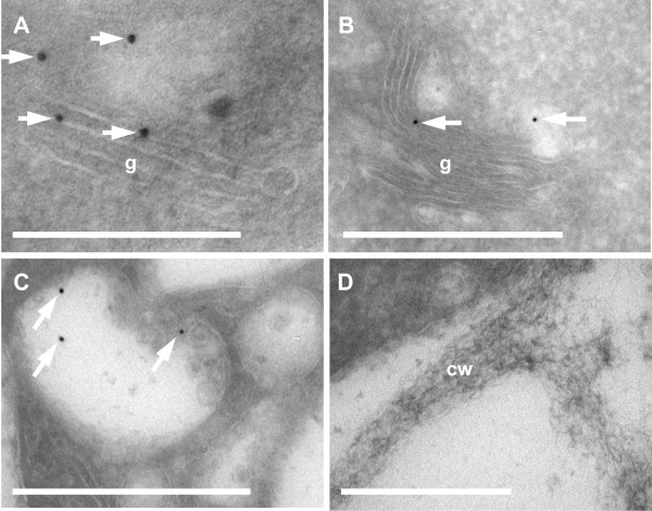
Figure 4.
Confirmation of the Golgi localization of AtAPY1 by immunogold labeling. (A-D) Transverse Tokuyasu-cryo-sections through root tips were imaged by TEM after imunogold labeling of AtAPY1-GFP using α-GFP antibodies (Torrey Pines) and Protein A 10-nm gold. Arrows mark gold particles. Independent experiments with Protein A 6-nm gold gave the same localization results presented here. Both approaches were repeated three times. Abbreviations: cw, cell wall, g, golgi stack, mvb, multivesicular body. Scale bars equal 200 nm in A and 500 nm in B-D.

