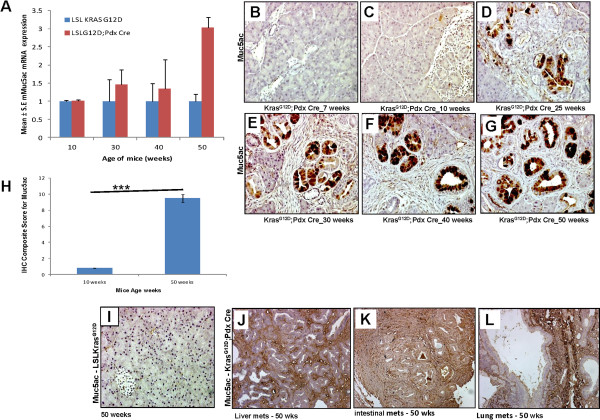
Figure 4.
Expression pattern of Muc5AC during the progression of pancreatic cancer in KrasG12Dmouse model. (A) Muc5AC mRNA expression was determined by quantitative Real Time PCR. (***p-value =0.0002) (B, C, D, E, F, and G) Muc5AC protein expression during the progression of pancreatic cancer in KrasG12D mouse model was analyzed by IHC. (H) Composite scores of pancreatic tissues of KrasG12D;Pdx1-Cre mice stained with Muc5AC antibody. (I) Normal pancreas from control mice (50 weeks) was stained negative for Muc5ac expression. (J, K, L) Muc5AC expression in the metastatic lesions involving liver, small intestines and lungs, respectively, in KrasG12D;Pdx1-Cre mice.

