
Figure 5.
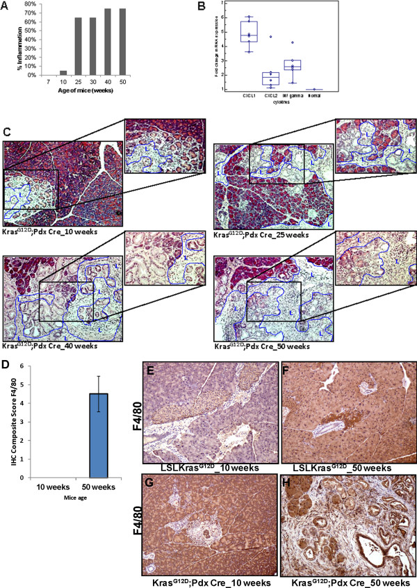
Inflammation during the progression of pancreatic cancer in KrasG12Dmouse model. (A) Percentage of inflammation in the pancreas of 7 to 50 weeks old KrasG12D;Pdx1-Cre mice evaluated on H&E stained tissue sections. (B) Expression of mRNA of the inflammatory cytokines/chemokines IFNγ, CXCL1 and CXCL2 in the pancreas of 50 weeks old KrasG12D;Pdx1-Cre mice compared to LSLKrasG12D (i.e. normal, unfloxed) animals. (C) Infiltration of lymphocytes into the pancreas of KrasG12D;Pdx1-Cre mice. Lymphocytes (L) that infiltrated into the pancreas of KrasG12D;Pdx1-Cre mice are shown within the blue boundaries in the H&E stained tissues. Light microscopic pictures are magnified 200x for each age group. D) Composite scores of pancreatic tissues of KrasG12D;Pdx1-Cre mice stained with F4/80 antibody for macrophages. (E, F, G, H) F4/80 marker expression for macrophages during the progression of pancreatic cancer in KrasG12D;Pdx1-Cre mouse model was analyzed by IHC.