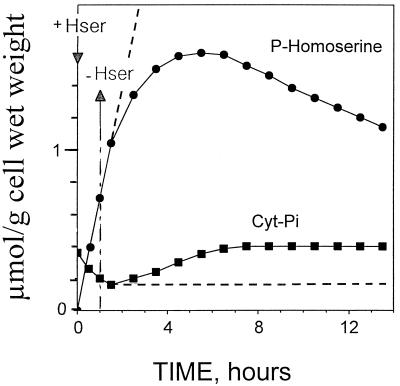
Figure 6.
Evolution of cytoplasmic Pi (Cyt-Pi) and phosphohomoserine in sycamore cells. In vivo 31P-NMR was used to determine Cyt-Pi and phosphohomoserine. At time 0, 100 μm homoserine was added to the perfusion medium at pH 6.0. After 1 h, cells were maintained in the medium (dashed lines) or perfused with a culture medium devoid of homoserine to remove extracellular homoserine (unbroken lines). The values, expressed as μmol g−1 cell wet weight, are from a representative experiment chosen from a series of five. Hser, Homoserine.

