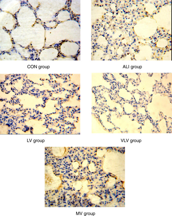
Figure 6.
eNOS protein in pulmonary tissues. The pulmonary tissues were analyzed for expression of eNOS protein by immunohistochemistry(IHC). Dark brown staining of indicates eNOS-positive cells. IHC stain of representative pulmonary artery endothelial cells from rats subjected to CON=Control Group, ALI=Acute lung injury Group, LV=Low tidal volume Group, VLV=Very low tidal volume Group, MV=Large tidal volume Group (magnification, ×400).

