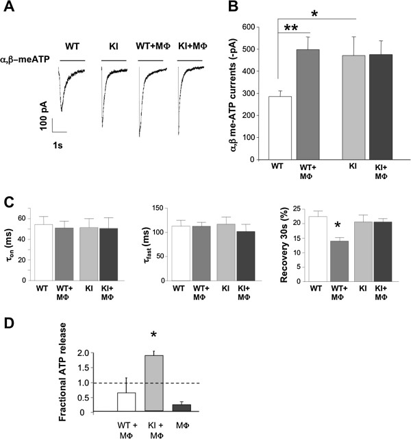
Figure 3.
Neuronal P2X3 receptor-mediated responses in basal conditions or in the presence of host macrophages. A, Representative examples of currents induced by application of α,β-meATP (10 μM, 2 s; horizontal bar) to trigeminal neurons from WT (n = 15 neurons) or R192Q KI (n = 17) cultures in standard conditions (left traces) or when co-cultured with macrophages (WT+MФ, .n = 24; KI+MФ, .n = 20. Note that macrophage co-culturing increases P2X3-mediated responses from WT neurons. Average data are plotted in B. * p < 0.05; ** p < 0.01. C, Rise time (left; expressed as time from 10 to 90% of peak amplitude), desensitization onset (middle; expressed as the first time constant, τfast, of current decay). Values are from 13-23 neurons. Recovery from desensitization (right; expressed as % of control amplitude in a paired pulse agonist application) was faster for WT+MФ vs WT; * p = 0.007. All responses were evoked by α,β-meATP (10 μM, 2 s). D, ATP medium content measured 5 h in WT or KI trigeminal neuron-macrophage co-cultures. Basal ATP levels present in culture macrophages are also shown. Data are expressed as fractional increase with respect to neuronal WT or KI cultures. n = 2, * p < 0.05.

