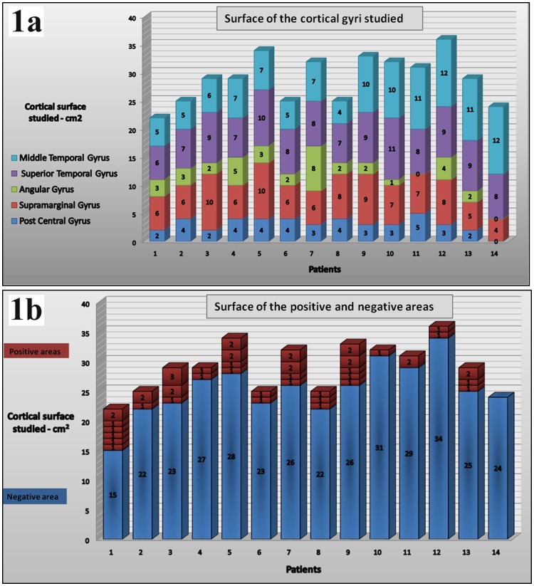
Figure 1. Number and size of reading interference areas.
Figure 1a shows in each patient the surface of the cortical gyri studied with some variations according to the tumor location and size of bone flap. Figure 1b illustrates the variability of the number and surface of the positive reading areas among this group of patient. A majority of positive areas were discrete (1 cm2) cortical areas; adjacent sites (2 cm2) found to be both involved in reading was observed 11 times, and only in a single case did we find a positive area of 3 cm2.

