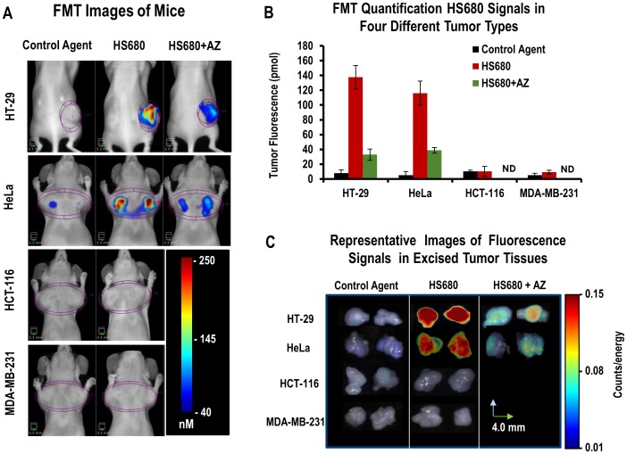
Figure 4. In vivo imaging of HS680 and control agent in mice bearing CA IX positive (HT-29 and HeLa) and CA IX negative (HCT-116 and MDA-MB-231) tumors, with and without AZ competition.
A, Representative FMT images and B, Tomographic quantitative analysis of tumor bearing mice injected with control agent and HS680 showing significant accumulations of HS680 fluorescence signals within HT-29 and HeLa tumors, but not in HCT-116 and MDA-MB-231 tumors, and less in the mice that were pre-injected with 10 mg/kg AZ (HS680+AZ) 1 hour before HS680 injection. Mice injected with control agents did not show any appreciable tumor fluorescence signals. HT-29 tumors are at the flank regions of the mice while all other tumor types are implanted in the mammary fat pads, and all tumors are housed with a 3D region of interest (ROI) in each image. ND indicates “Not determined.” C, Representative FRI images of four types of tumors collected following the FMT imaging, showing ex-vivo validation of in vivo imaging results.

