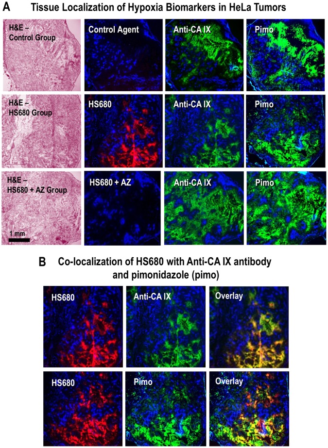
Figure 6. Localization of HS680 and tumor hypoxia markers in HeLa xenografts.
A, The tissue staining patterns of the control agent, HS680, and HS680+AZ (red) from the same or adjacent tumor sections as staining with fluorescent CA IX antibody or pimonidazole (green) and the Hoechst perfusion stain (blue). H&E staining of tissue sections that were used for localization are shown on the left side of images. HS680 was specifically localized in regions with low Hoechst staining indicative of low oxygen (less perfused) but positive staining with both the CA IX antibody and Pimonidazole. Preinjection of the mice with unlabeled AZ blocked the binding of HS680 to control levels. B, Co-localization (overlay) of HS680 with CA IX antibody or pimonidazole (hypoxyprobe) is shown on the right side images indicating HS680 was clearly co-localized with both anti-CA IX antibody and hypoxyprobe (pimonidazole). Similar results were obtained using HT-29 tumors (Figure S4A and Figure S4B).

