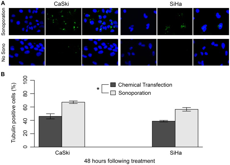
Figure 3. Detection of 4 µg/mL tubulin antibody within CaSki and SiHa cells.
(A) Immunofluorescence detection of the tubulin antibody 48 hours following exposure of the cells to sonoporation or sham no HIFU treatment. Nuclei were counterstained blue with DAPI (left photo). Green signal indicates internalized tubulin antibody detected by the application of a secondary fluorophore (centre photo). Images merged (right photo). Cells were visualized using 400× magnification. (B) Tubulin antibody positive cells (%) 48 hours following both chemical transfection and sonoporation with 4 µg/mL antibody. Sonoporation resulted in a higher antibody transfection efficiency than that obtained with the traditional chemical reagent, HiPerFect (P<0.001, n = 3). Statistical analysis was performed using a two-way ANOVA with Student’s two-sample t-tests post hoc. Error bars represent mean +/− SEM. “*” denotes significance.

