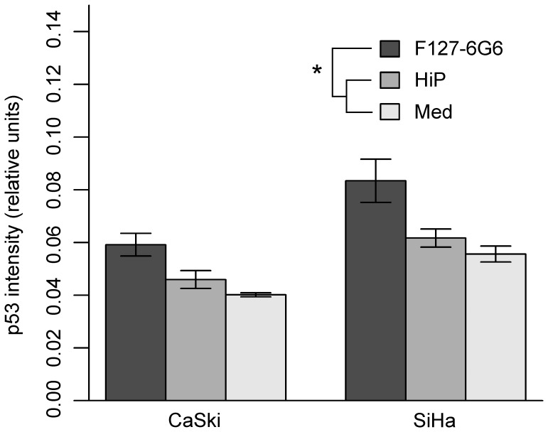
Figure 6. Average quantity of p53 per cell following chemical transfection with 40 µg/mL F127-6G6 E6 antibody.
p53 quantity is evidenced by intensity of p53 staining. Cells transfected with the antibody show a higher quantity of p53 per cell (i.e. a higher intensity of p53 staining) after 48 hours than those in control wells treated with only the HiPerFect (HiP) transfection reagent or culture medium (Med) alone (P = 0.005, P<0.001, n = 3 for both). Also, SiHa cells showed a greater quantity of p53 per cell than CaSki (P<0.001, n = 3). Statistical analysis was performed using a two-way ANOVA followed by Tukey HSD contrast tests post hoc. Error bars represent mean +/− SEM. “*” denotes significance.

