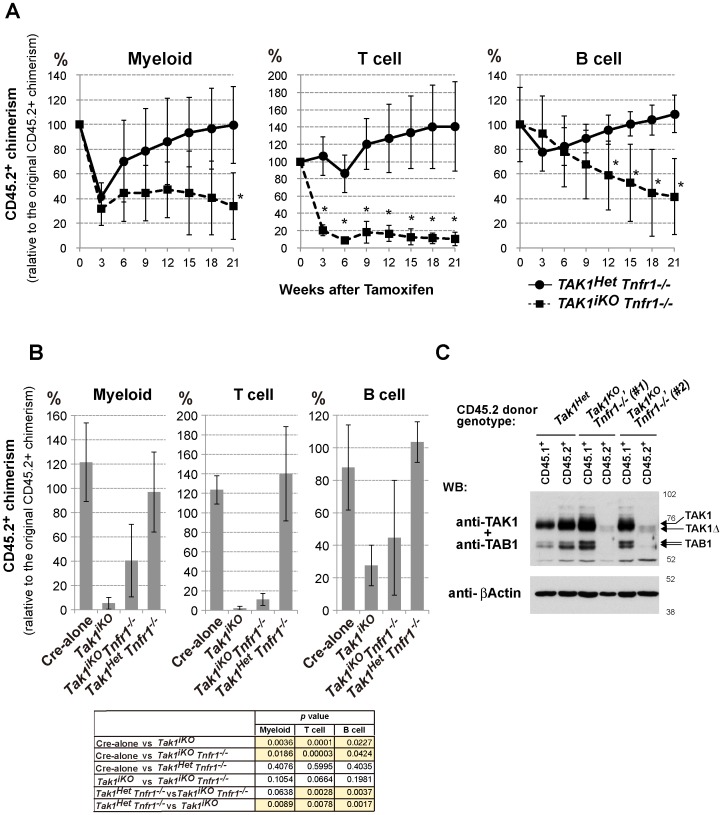
Figure 5. Ablation of TNF signaling partially restores the reconstitution potential of Tak1-deficient HSCs.
(A) Competitive reconstitution assay. 2×10⁵ BMN cells from control Tak1Het Tnfr1−/− (n = 3) or Tak1iKO Tnfr1−/− mice (n = 4) (CD45.2+) were transplanted into lethally irradiated recipients (CD45.1+) together with 2×10⁵ competitor wild type BMN cells (CD45.1+). At six weeks post transplantation (designated as Week 0), the chimerism of myeloid, T and B cells in the recipients’ PB was analyzed, and then the recipients were i.p. injected with tamoxifen at 160 mg/kg body weight for three consecutive days. The chimerism of PB cells was monitored every three weeks, and is shown as the mean ± S.D. (*p<0.05) (B) Competitive reconstitution assay of Cre-alone (n = 3) and Tak1iKO (n = 3) was also performed as described above, and compared with Tak1Het Tnfr1−/− (n = 3) and Tak1iKO Tnfr1−/− (n = 4) at 18 weeks post-tamoxifen injection. The table shown below the graphs indicates statistical significance for the indicated comparisons. P values of less than 0.05 are highlighted. (C) Expression of TAK1 and TAB1 proteins in the donor-derived splenocytes. Whole spleen cells from control Tak1Het and two independent Tak1iKO Tnfr1−/− transplanted mice (#1 and #2) at 15 weeks post-tamoxifen injection were sorted into the CD45.1+ or CD45.2+ population. Total cell lysates from the sorted splenocytes from control Tak1Het, and Tak1iKO Tnfr1−/− #1 and #2 were analyzed by SDS-PAGE and Western blotted with anti-TAK1, TAB1 or anti-β-actin antibodies. The positions of molecular weight markers are shown on the right. The arrows indicate the bands corresponding to endogenous TAK1 and TAB1, and truncated TAK1 (TAK1Δ) resulting from Cre-mediated recombination.

