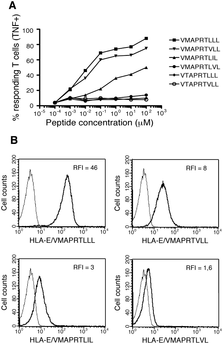
Figure 2. Characterization of CMV/HLA-I-derived peptides recognized by HLA-E-restricted CD8 T cells.
A/TNF production in response to stimulation with.221 cells pulsed with synthetic peptides.221 cells were incubated for 1 h with range concentrations of the indicated peptides before addition of MART.22 T cells. After 6 h, T cells were fixed, permeabilized and stained for intracellular TNF-α. Results are expressed as percentage of TNF-producing T cells. B/Peptide-MHC tetramer staining of HLA-E-restricted CD8 T cells. MART.22 T cells were incubated for 1 h with biotyniled HLA-E monomers refolded with the indicated peptides and tetramerized with PE-coupled streptavidin. Peptide-HLA-E tetramers staining was assessed by flow cytometry and RFI are indicated.

