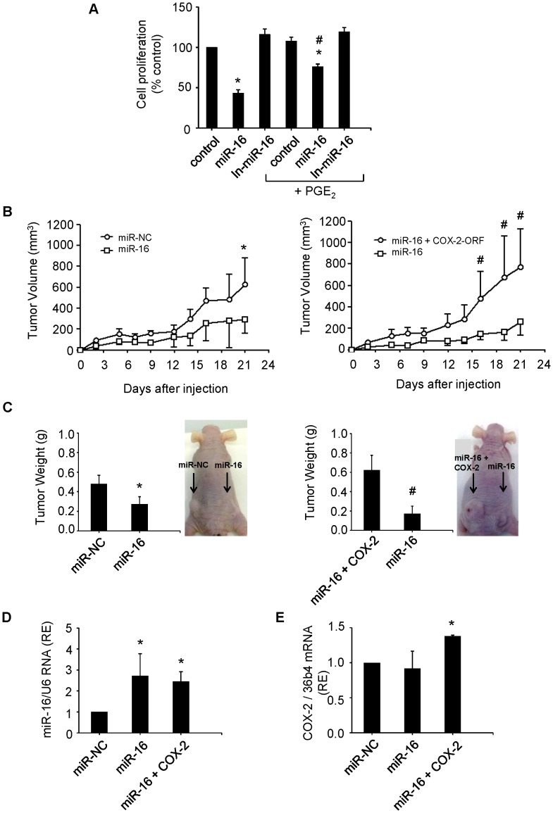
Figure 7. miR-16 suppresses growth of hepatoma cells in vitro and tumorigenicity in vivo.
(A) Hep3B cells were transfected with 50 nM miR-16 or In-miR-16. The effect on cell proliferation was determined by MTT assay at 48 h after transfection. *p< 0.05 vs. the control and # p< 0.05 vs. the miR-16 transfection condition. (B) Tumor growth curves measured after subcutaneous injection of WRL68 cells transiently transfected with miR-16, miR-NC or miR-16 with a hCOX-2 expression vector lacking 3′ UTR (miR-16+COX-2). Tumor volume (V) was monitored by measuring the length (L) and width (W) with calipers and calculated with the formula (L×W2)×0.5. Tumor growth was measured every 2–3 days. *p< 0.05 vs. the miR-NC and # p<0.05 vs. miR-16 (C) Tumor weight and a representative picture of the tumors. At 21 days after injection, mice were killed and tumors were weighed after necropsy. *p< 0.05 vs. the miR-NC or # vs. the miR-16+ COX-2 condition. (D–E) miR-16 and human COX-2 expression in tumors using real-time PCR normalized against U6 RNA levels and refers to miR-NC as 1 or 36b4 mRNA, respectively. *p< 0.05 vs. the miR-NC Data are reported as means±SD of three independent experiments or five animals per condition.

