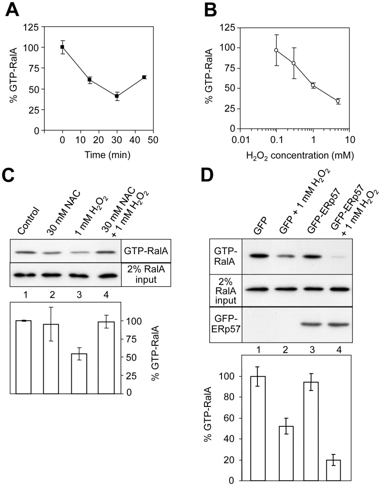
Figure 5. Oxidative stress inhibits cellular RalA activity via ERp57.
GST-RalBD Ral activity assays monitoring the level of GTP-bound endogenous RalA (A) A431 cells were treated with 2 mM H2O2 and then the amount of active RalA-GTP was measured over 45 min. Error bars show SEM (n = 3). (B) Serum starved A431 cells were treated with various concentrations of H2O2 for 30 minutes and then RalA-GTP was measured (n = 3). (C) RalA activity assays examining the effect of pre-treatment with the antioxidant N-acetylcysteine (NAC) for 18 hours. The intensity of RalA staining in the upper panel indicates the level of RalA-GTP in a representative experiment, and the quantification of these bands by densitometry is shown beneath (n = 6). The relative levels of total cellular RalA are shown in the lower panel (from 2% of total protein loaded onto the GST-RalBD column). (D) Expression of GFP-ERp57 enhances the H2O2-induced inactivation of RalA in A431 cells. In the lower panel, error bars represent SEM (n = 9).

