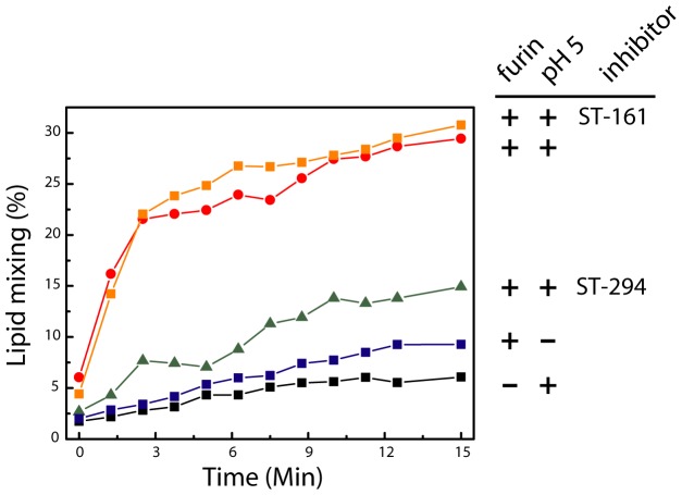
Figure 6. pH-induced membrane fusion by rGPCfur proteoliposomes: lipid mixing.
rGPCfur was incorporated into POPG-POPC liposomes and mixed with target POPG-POPC liposomes doped with 1% Rhodamine-PE. In most cases, the rGPCfur proteoliposomes were first treated with sFurin (as indicated by+in the first position of the labels, at right). Exposure to acidic pH at the start of the experiment (time = 0) is indicated by+in the second position of the labels. 15 µM of ST-294 or ST-161 was present prior to and during exposure to acidic pH, where indicated. Lipid mixing and the resulting dequenching of the rhodamine fluorophore were measured at 600 nm (excitation 508 nm). Complete dequenching (100%) was determined by subsequent solubilization in Triton X-100 nonionic detergent.

