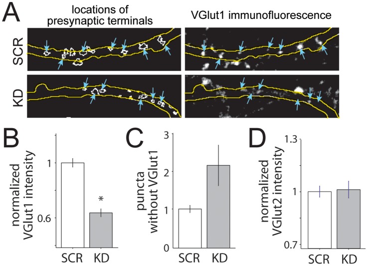
Figure 6. Transfection with VGLUT1 shRNA results in significant knockdown of VGLUT1 at presynaptic terminals.
(A) Images of VGLUT1 presynaptic puncta near and within axons of cultured neurons transfected with control non-targeting shRNA (SCR, top) and VGLUT1-targeting shRNA (KD, bottom). Transfected neurons expressed GFP throughout the cell, and axons were identifiable by fluorescence and morphological characteristics (outlined in yellow). Synaptophysin-expressing presynaptic terminals were selected automatically via an imaging processing algorithm and identified as regions of interest (outlined in white, left). VGLUT1 immunofluorescence was measured within each region of interest. Presynaptic terminals within transfected axons are indicated with blue arrows (left and right). (B) The mean intensity of VGLUT1 decreased significantly in KD puncta. *, p<0.001. (C) The percentage of puncta without VGLUT1 increased for the KD group (n = 1305 and 978 SCR and KD puncta, respectively, from 3 independent experiments; data are normalized to SCR). (D) VGLUT2 was not up-regulated in KD neurons (p = 0.8357, n = 440 and 246 puncta from 11 SCR and 14 KD axons, respectively).

