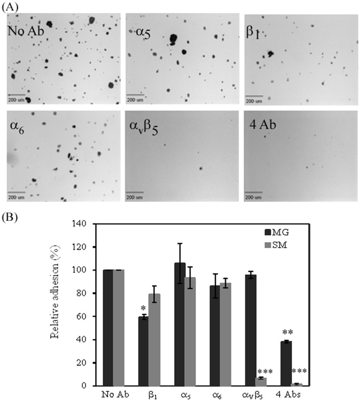
Figure 3. The role of integrin receptors in hiPSC adhesion to Synthemax Surface and Matrigel.
hiPSCs were incubated with indicated anti-integrin antibodies before seeding onto the corresponding surface. (A) Micrographic images of cell attachment to the Synthemax Surface after integrin treatment. Scale bar: 200 µm. (B) Relative cell adhesion determined at 1 h post seeding. A cell adhesion was estimated by counting the number of colonies in 14 randomly selected fields. The relative cell adhesion was normalized to that for integrin-untreated cells. All results were expressed as the mean ± SD (n = 14) in two typical measurements of more than 3 independent experiments. *: p = 0.0093; **: p = 0.0002; ***: p<2.75E-10. Symbols: Ab, antibody; MG, Matrigel; SM, Synthemax Surface.

